


Welcome to Fantastic Voyage, where we explore the brain one region at a time. (New here? Start with What’s Hiding Inside That Three-Pound Lump Between Your Ears? for the big picture, or jump right in.)
In 2003, a man known in the medical literature as TN suffered two strokes five weeks apart. The first destroyed the visual cortex on the left side of his brain. The second destroyed it on the right. His eyes were perfectly healthy, but the part of the brain that turns what the eyes detect into a conscious picture had been wiped out on both sides. TN was completely blind.
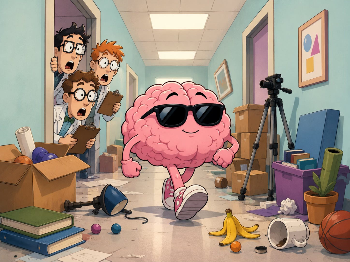
Five years later, neuroscientist Beatrice de Gelder arranged an experiment. Her team lined a long corridor with obstacles, everything from boxes to a camera tripod. They told TN the hallway was empty and asked him to walk without his cane. A researcher followed close behind in case he stumbled.
He never stumbled. TN weaved between obstacles, sidestepping a box here and angling around the tripod there, never touching a single object. When he reached the end of the corridor, the assembled researchers broke into spontaneous applause. See the original footage below.
TN navigating cluttered hallway. Curr Biol. 2008;18(24):R1128-R1129.
TN had no idea why they were clapping. He did not know the obstacles were there. He did not know he had avoided them.
Your Brain’s Movie Screen
The region TN lost is called the primary visual cortex, or V1. It sits at the very back of the brain, in the occipital lobe, as far from the eyes as you can get. Light enters your eyes at the front of your head and the signal travels all the way to the back before you actually see anything.
Under a microscope, a prominent stripe of nerve fibers runs through V1, earning it the nickname striate cortex. (Striate means “striped.”) Two neuroscientists, David Hubel and Torsten Wiesel, spent decades mapping what its cells do, work that earned them the Nobel Prize in 1981.
What they found is that V1 does not receive a photograph from the eyes. It takes the raw visual signal apart. Individual neurons respond only to specific features. Some fire when they detect an edge tilted at one angle. Others respond to a different angle. These cells are organized into orientation columns, tiny vertical stacks that share the same preferred tilt. Rotate the stimulus a few degrees and the neighboring column takes over.
V1 also contains zones called blobs (yes, that is the official term) where neurons analyze color, and ocular dominance columns, alternating strips of tissue that keep input from your left and right eyes separate so the brain can calculate depth. Each square millimeter works as a processing module that Hubel and Wiesel called a hypercolumn, handling everything from edge detection to depth perception for one small patch of the visual world.
From V1, information splits into two highways. One runs upward toward the parietal lobe, the “where” pathway we visited in What Happens When Your Brain Deletes an Entire Side of Reality?, tracking where objects are in space. The other runs downward toward the temporal lobe, the “what” pathway, figuring out what you are looking at.
Seeing Without Knowing
If V1 is so critical, how did TN navigate that hallway?
V1 is not the only part of the brain that receives visual information. Some nerve fibers from the eyes bypass the cortical route entirely and go straight to an older structure called the superior colliculus, a small mound in the midbrain that evolved long before the cortex existed. The superior colliculus handles rapid, unconscious visual responses. It is the reason you flinch when something flies toward your face before you consciously register what it is.
In 1974, Oxford neuroscientist Lawrence Weiskrantz tested a patient called DB, whose right V1 had been surgically removed. In forced-choice experiments, DB could point to objects and detect motion in his blind field, performing far above chance. But he insisted he saw nothing and believed he was guessing. Weiskrantz coined the term “blindsight.” DB’s eyes were sending information through pathways that bypassed V1, and his brain was using it to guide behavior without ever producing a conscious experience of seeing.
TN’s case, published in 2008, was the most dramatic confirmation. He had lost V1 in both hemispheres, not just one. Yet the ancient subcortical visual system, the one that predates the cortex by hundreds of millions of years of evolution, still worked.
Your brain does not have a single vision system. It has at least two, running in parallel. One builds the conscious, full-color experience you call seeing. The other operates below awareness, keeping you safe even when the conscious system goes dark.
Next, we travel inward to a seahorse-shaped structure buried deep in the temporal lobe. When it was removed from a single patient in 1953, the result changed everything neuroscience thought it knew about how memories are made.
References
Kandel ER, Schwartz JH, Jessell TM. Principles of Neural Science, 4th ed. McGraw-Hill, 2000. Chapters 25, 27, and 28.
Weiskrantz L, Warrington EK, Sanders MD, Marshall J. Visual capacity in the hemianopic field following a restricted occipital ablation. Brain. 1974;97(4):709-728. doi:10.1093/brain/97.1.709
de Gelder B, Tamietto M, van Boxtel G, et al. Intact navigation skills after bilateral loss of striate cortex. Curr Biol. 2008;18(24):R1128-R1129. doi:10.1016/j.cub.2008.11.002
Hubel DH, Wiesel TN. Receptive fields, binocular interaction and functional architecture in the cat’s visual cortex. J Physiol (Lond), 1962; 160:106-154.